Avatar

Tick Season: Protect Your Pet!

Learn More
Search for AREN


Diagnostic Imaging for Pets

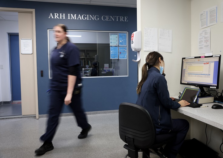
Diagnostic imaging is the bedrock to any veterinary practice, allowing specialists to diagnose your pet’s diseases, plan treatments, and guide treatment procedures. At the Animal Referral & Emergency Network we're fortunate to have some of the most advanced imaging equipment in Australia. This allows our specialists to offer the highest standard of care for your loved pet.


We offer the following services:

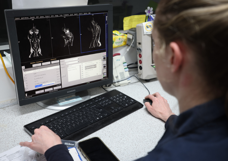
  • Magnetic Resonance Imaging (MRI)
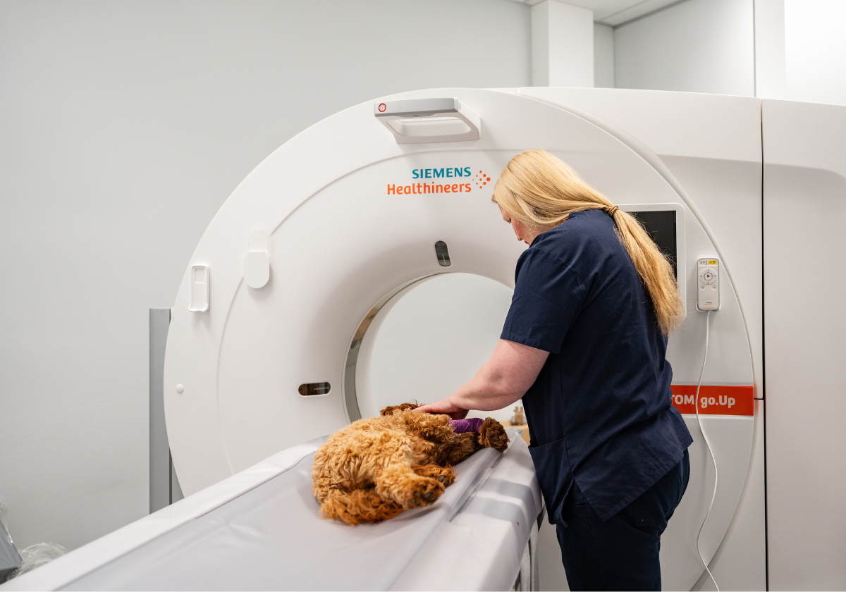
  • Computed Tomography (CT)
  • Fluoroscopy
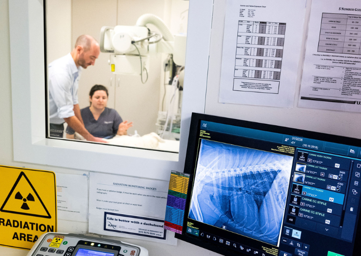
  • General X-ray
  • Ultrasound


We take pride in our specialty services

Magnetic resonance imaging (MRI) uses a strong magnetic field and radiofrequencies to acquire high-resolution cross-sectional images of the body. There is no ionising radiation involved with MRI studies and it is a non-invasive imaging modality, reducing the potential complications that may occur with traditional myelography. MRI is useful in the diagnosis of acute and chronic spinal pathology, brain lesion and musculoskeletal disease as opposed to the traditional imaging methods as it provides superior soft tissue definition of these regions that CT or X-rays do not permit.

Our examinations are comparable to those you would find in the human world, only all our patients require general anaesthetic to remain still for the scan. Due to the complex nature of MRI, these studies typically take longer than a CT. At VRH we have support from both specialist anaesthetists and a full-time anaesthetic nurse, dedicated to managing the anaesthesia and safety of your pet.

Most of our MRI cases are neurological studies, and include assessing our patients for causes of seizures, changes in mentation, spinal pain, nerve deficits and paralysis. However, we also scan for musculoskeletal diseases in the shoulders (like biceps tendons or medial glenohumeral ligament instability), stifle (like meniscal damage) and elbows.

MAKE A REFERRAL

Computed tomography (CT) is also called CAT scan, is a non-invasive diagnostic imaging procedure that allows detailed imaging of all parts of the body. CT uses ionising radiation (x-rays) to create high-resolution cross-sectional images of the body, showing detailed information not seen on conventional x-rays. CT allows us to visualise diseases not seen on ultrasound and far more sensitive for masses than x-rays. 3D reformatting capabilities of CT allow surgical planning, 3D templating and creation of patient specific surgical implants and drill-guides. Contrast (dye) is typically used to provide better differentiation of tissues and structures in the body.

Due to its fast nature we can images areas such as the head, neck, chest, abdomen and pelvis in a minute. This is useful with trauma cases as CT provides superior bony detail compared to other imaging modalities and can provide detailed information on complex fracture. It guides surgeons what areas to focus on rather than having to have exploratory surgery. CT is an excellent tool for localisation of lesions, especially in the nose, abdomen and thorax that can then be sampled under CT or ultrasound guidance.

MAKE A REFERRAL

Fluoroscopy allows us to view and acquire real time images using x-rays during interventional and surgical procedures. In surgery, fluoroscopy allows our surgeons to place and assess surgical implants in real time, using minimally invasive approaches, and not having to revise the surgery after x-rays are taken. Fluoroscopy also allows us to assess motility and function of organs such as the trachea for collapse, or the oesophagus for swallowing function. Fluoroscopy is also used to perform minimally invasive procedures, such as placing stents or assessing patency of organs, such as the ureters. Thus, fluoroscopy compliments both our surgical and medicine departments.

MAKE A REFERRAL

X-ray will always be a great imaging tool, which is readily available and cost effective. It can diagnose gross pathology, acute injury, follow up assessment and to monitor for changes. X-rays are the main imaging modality used by all our departments, and is performed by our well-trained veterinarians, nurse-technicians and supported by radiographer team.

MAKE A REFERRAL

Ultrasound is an excellent, painless and safe diagnostic imaging modality. It can be done without general anaesthetic, but uses sedation to keep your pet calm whilst on the ultrasound table or during sampling. Ultrasound images are obtained by using sound waves transmitted through and reflected by the organs of the body. Ultrasound allows us to assess the organs of the abdomen, thoracic wall, eyes, muscles, tendons, ligaments and joints. At VRH we can also perform basic echocardiography, from which we can diagnosis most structural diseases of the heart and then correspond with a cardiologist for further diagnosis and treatment.

Our ultrasounds are performed by specialist-trained ultrasonographer. The ultrasound study usually takes 45minutes, where a scan can be performed, and sampling done with direct communication with of your treating medicine, emergency and critical care, or surgical specialist.

MAKE A REFERRAL


How AREN’s specialist Diagnostic Imaging can help

Advanced imaging for precise diagnosis. Our state-of-the-art technology and expert team deliver accurate insights, supporting timely and effective treatment for your pet’s health needs.


Our Network

Our Animal Referral & Emergency Network is the largest specialty and referral network in Australia, consisting of more than 20 sites. With more than 1,200 dedicated team members, including approximately 600 nurses and almost 400 veterinarians (including specialists and registrars), we provide exceptional care for your pets. Count on us for expert medical attention and comprehensive veterinary services.