Discussion
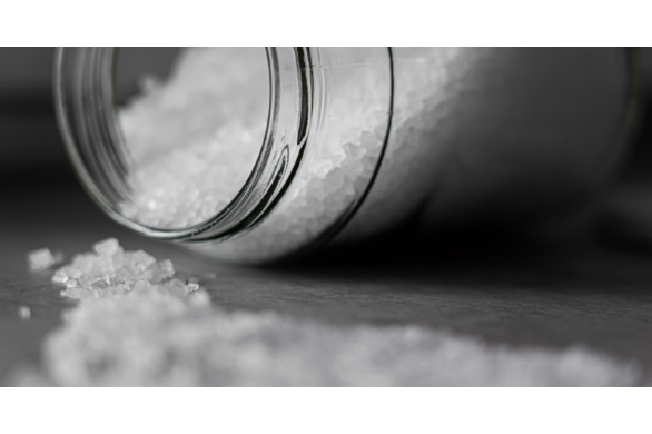
Hypernatraemia in dogs and other animals can be caused by a sodium gain or free water loss. Where a sodium gain is present, the cause is often easily identified such as salt ingestion or administration of hypertonic saline or sodium bicarbonate. Free water losses can be more elusive and include inadequate access to water, primary hypodipsia, diabetes insipidus, fever, renal losses, gastrointestinal losses, third spacing, etc. In this case, our patient had consumed large amounts of salt water, leading to a spike in their sodium levels.
A helpful way of approaching a patient who has hypernatremia is to assess their volume status (Figure 1). Animals with sodium gain are often hypervolaemic. Animals with pure free water loss (e.g., diabetes insipidus, inadequate access to water) are normovolaemic. Those with hypotonic losses (water loss in excess of electrolyte loss) are usually hypovolaemic and include gastrointestinal and renal losses and third spacing.
In cases of mild to moderate hypernatraemia, there may be no clinical signs, especially if the rise in sodium has occurred over many days. However, once plasma sodium levels increase >170 mmol/L, neurological signs such as obtundation, muscular weakness, behavioral changes, disorientation, ataxia, seizures, and coma may occur.
Sodium is the main determinant of plasma osmolality. When there is excessive sodium in the extracellular fluid compartment it becomes hyperosmolar, and water moves from the relatively hypo-osmolar intracellular fluid into the extracellular fluid causing cell shrinkage. The brain can protect against neuronal water loss initially with fluid and ionic shifts and accumulation of organic solutes to prevent large volume loss from the neurons. However, rapid or severe changes to cell volume cannot easily be compensated for. Neurones are the cell type least tolerant to changes in volume, resulting in a predominance of neurological signs in disorders of sodium balance.
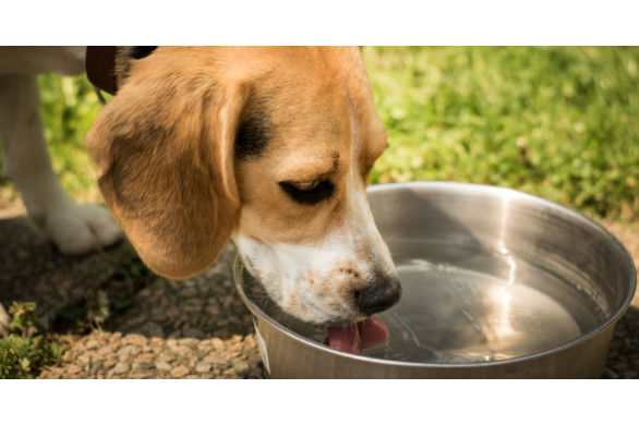
Regardless of the cause, all cases of hypernatraemia are considered to have a free water deficit. Treatment of hypernatraemia involves correcting the free water deficit by providing the animal with water replacement. In mild cases, oral intake of water can be encouraged. However, in more severe cases, intravenous fluid therapy provides less margin for error in controlling the sodium descent. The intravenous fluid of choice is 5% dextrose as free water is the carrier and water is produced through glucose metabolism.
An animal’s free water deficit is calculated using the following formula: Free water deficit (L) = {(current [Na+] ÷ normal [Na+]) – 1} x (0.6 x body weight in kg).
The number of hours over which to deliver this volume is calculated based on the recommended limitation of a drop in sodium no faster than 1.0 mmol/L/h. This prevents the rapid redistribution of water across the cell membrane into the intracellular space, which results in cellular swelling.
In the case above the free water deficit was calculated as [(180/144.5)-1] x (0.6 x 18) = 0.246 x 10.8 = 2.66 L.
To return to a normal sodium of 144.5 mmol/L, the plasma sodium in this animal would need to reduce by at least 35.5 mmol/L. Therefore, to reduce the sodium by a rate no faster than 1 mmol/L/h, the free water volume (2.66 L) would need to be administered over at least 35.5 hours. Therefore, the fluid rate of 5% dextrose to be administered would be 2660 ml/35.5 h = 75 mL/h.
Plasma sodium concentration should be measured at least every 4 hours while sodium levels are being corrected. The rate of free water replacement may need to be adjusted if the sodium concentration is decreasing more rapidly than 1 mmol/L/h.
Free water replacement leads to movement of water into cells. If this replacement occurs too rapidly, cells will swell. Neurones are particularly sensitive to this water intake and cerebral oedema can rapidly develop. The clinical signs of cerebral oedema are similar to those of hypernatraemia including obtundation, head pressing, seizures, coma, and changes in behaviour or movement. Cerebral oedema should be suspected when a symptomatic hypernatraemic animal initially improves with free water replacement and then deteriorates neurologically, or an asymptomatic hypernatraemic animal receiving treatment develops neurological signs.
If any of these symptoms develop during treatment, intravenous fluids or oral water intake should be ceased. Plasma sodium concentration should be measured to demonstrate a rapid decrease and treatment for cerebral oedema should be initiated. Treatment of cerebral oedema includes either a single dose of mannitol at 0.5-1 g/kg intravenously over 20 to 30 minutes or judicious administration of hypertonic saline.
References:
DiBartola, SP. Fluid, Electrolyte and Acid-Base Disorders in Small Animal Practice. Elsevier 2012.
Silverstein DC, Hopper K. Small Animal Critical Care Medicine, 2nd Ed. Elsevier 2014.